Fallen Arches Explained
Overview
It is rare to find someone who walks with both feet in perfect alignment. Often we walk on the inside or outsides of our feet, or with our toes or heels rotated inward. These typical walking patterns can cause many problems not just with the feet but also for the whole body as the alignment of the feet sets the foundation for the whole body?s alignment. One of the most common effects of improper alignment is known as flatfoot, or fallen arches.
Causes
Some people develop fallen arches because they tend to pronate, or roll inwards on the ankles, says the Instep Foot Clinic. Other people may simply have under-developed muscles in their arches. Your arches help your feet bear weight and are supported in this job by muscles and tendons in your feet and ankles. So, while fallen arches aren?t usually serious, they can cause pain in your feet, ankles, knees and/or hips due to your reduced weight-bearing ability. In these cases, treatment may be required. Orthotics that sit in your shoes and support your arches are a common solution, as are exercises to strengthen and stretch your feet and leg muscles.
Symptoms
Symptoms that should be checked by a pediatrician include foot pain, sores or pressure areas on the inner side of the foot, a stiff foot, limited side-to-side foot motion, or limited up-and-down ankle motion. For further treatment you should see a pediatric orthopedic surgeon or podiatrist experienced in childhood foot conditions.
Diagnosis
If your child has flatfeet, his or her doctor will ask about any family history of flatfeet or inherited foot problems. In a person of any age, the doctor will ask about occupational and recreational activities, previous foot trauma or foot surgery and the type of shoes worn. The doctor will examine your shoes to check for signs of excessive wear. Worn shoes often provide valuable clues to gait problems and poor bone alignment. The doctor will ask you to walk barefoot to evaluate the arches of the feet, to check for out-toeing and to look for other signs of poor foot mechanics. The doctor will examine your feet for foot flexibility and range of motion and feel for any tenderness or bony abnormalities. Depending on the results of this physical examination, foot X-rays may be recommended. X-rays are always performed in a young child with rigid flatfeet and in an adult with acquired flatfeet due to trauma.
arch support plantar fasciitis
Non Surgical Treatment
The type of treatment will depend on the stage of PTTD present. There are four stages of posterior tibial tendon dysfunction. Stage I. The posterior tibial tendon is inflamed but has normal strength. There is little to no change in the arch of the foot. The patient can still perform a single-limb heel rise and has a flexible hindfoot. Orthotic treatment options include modified off the shelf inserts and custom molded orthotics. Stage 2. The tendon is partially torn or shows degenerative changes and as a result loses strength.There is considerable flattening of the arch without arthritic changes in the foot. The patient cannot perform single-limb heel rise. Pain is now present on the lateral aspect of the ankle. Orthotic treatment is similar as that in stage I, with the addition of more rigid arch supports and wedging. Stage 3. Results when the posterior tibial tendon is torn and not functioning. As a result the arch is completely collapsed with arthritic changes in the foot. A solid ankle AFO is suggested in conjunction with a modified orthopedic shoe. Stage 4. Is identical to stage three except that the ankle joint also becomes arthritic. A rigid AFO and modified orthopedic shoe is required.
Surgical Treatment

A better approach is to strengthen the weakened ligaments with Prolotherapy, supplemented by an arch support if the condition has existed for several years. Chronic pain is most commonly due to tendon and ligament weakness, or cartilage deterioration. The safest and most effective natural medicine treatment for repairing tendon, ligament and cartilage damage is Prolotherapy. In simple terms, Prolotherapy stimulates the body to repair painful areas. It does so by inducing a mild inflammatory reaction in the weakened ligaments and cartilage. Since the body heals by inflammation, Prolotherapy stimulates healing. Prolotherapy offers the most curative results in treating chronic pain. It effectively eliminates pain because it attacks the source: the fibro-osseous junction, an area rich in sensory nerves. What?s more, the tissue strengthening and pain relief stimulated by Prolotherapy is permanent.
After Care
Time off work depends on the type of work as well as the surgical procedures performed. . A patient will be required to be non-weight bearing in a cast or splint and use crutches for four to twelve weeks. Usually a patient can return to work in one to two weeks if they are able to work while seated. If a person's job requires standing and walking, return to work may take several weeks. Complete recovery may take six months to a full year. Complications can occur as with all surgeries, but are minimized by strictly following your surgeon's post-operative instructions. The main complications include infection, bone that is slow to heal or does not heal, progression or reoccurrence of deformity, a stiff foot, and the need for further surgery. Many of the above complications can be avoided by only putting weight on the operative foot when allowed by your surgeon.
It is rare to find someone who walks with both feet in perfect alignment. Often we walk on the inside or outsides of our feet, or with our toes or heels rotated inward. These typical walking patterns can cause many problems not just with the feet but also for the whole body as the alignment of the feet sets the foundation for the whole body?s alignment. One of the most common effects of improper alignment is known as flatfoot, or fallen arches.
Causes
Some people develop fallen arches because they tend to pronate, or roll inwards on the ankles, says the Instep Foot Clinic. Other people may simply have under-developed muscles in their arches. Your arches help your feet bear weight and are supported in this job by muscles and tendons in your feet and ankles. So, while fallen arches aren?t usually serious, they can cause pain in your feet, ankles, knees and/or hips due to your reduced weight-bearing ability. In these cases, treatment may be required. Orthotics that sit in your shoes and support your arches are a common solution, as are exercises to strengthen and stretch your feet and leg muscles.
Symptoms
Symptoms that should be checked by a pediatrician include foot pain, sores or pressure areas on the inner side of the foot, a stiff foot, limited side-to-side foot motion, or limited up-and-down ankle motion. For further treatment you should see a pediatric orthopedic surgeon or podiatrist experienced in childhood foot conditions.
Diagnosis
If your child has flatfeet, his or her doctor will ask about any family history of flatfeet or inherited foot problems. In a person of any age, the doctor will ask about occupational and recreational activities, previous foot trauma or foot surgery and the type of shoes worn. The doctor will examine your shoes to check for signs of excessive wear. Worn shoes often provide valuable clues to gait problems and poor bone alignment. The doctor will ask you to walk barefoot to evaluate the arches of the feet, to check for out-toeing and to look for other signs of poor foot mechanics. The doctor will examine your feet for foot flexibility and range of motion and feel for any tenderness or bony abnormalities. Depending on the results of this physical examination, foot X-rays may be recommended. X-rays are always performed in a young child with rigid flatfeet and in an adult with acquired flatfeet due to trauma.
arch support plantar fasciitis
Non Surgical Treatment
The type of treatment will depend on the stage of PTTD present. There are four stages of posterior tibial tendon dysfunction. Stage I. The posterior tibial tendon is inflamed but has normal strength. There is little to no change in the arch of the foot. The patient can still perform a single-limb heel rise and has a flexible hindfoot. Orthotic treatment options include modified off the shelf inserts and custom molded orthotics. Stage 2. The tendon is partially torn or shows degenerative changes and as a result loses strength.There is considerable flattening of the arch without arthritic changes in the foot. The patient cannot perform single-limb heel rise. Pain is now present on the lateral aspect of the ankle. Orthotic treatment is similar as that in stage I, with the addition of more rigid arch supports and wedging. Stage 3. Results when the posterior tibial tendon is torn and not functioning. As a result the arch is completely collapsed with arthritic changes in the foot. A solid ankle AFO is suggested in conjunction with a modified orthopedic shoe. Stage 4. Is identical to stage three except that the ankle joint also becomes arthritic. A rigid AFO and modified orthopedic shoe is required.
Surgical Treatment
A better approach is to strengthen the weakened ligaments with Prolotherapy, supplemented by an arch support if the condition has existed for several years. Chronic pain is most commonly due to tendon and ligament weakness, or cartilage deterioration. The safest and most effective natural medicine treatment for repairing tendon, ligament and cartilage damage is Prolotherapy. In simple terms, Prolotherapy stimulates the body to repair painful areas. It does so by inducing a mild inflammatory reaction in the weakened ligaments and cartilage. Since the body heals by inflammation, Prolotherapy stimulates healing. Prolotherapy offers the most curative results in treating chronic pain. It effectively eliminates pain because it attacks the source: the fibro-osseous junction, an area rich in sensory nerves. What?s more, the tissue strengthening and pain relief stimulated by Prolotherapy is permanent.
After Care
Time off work depends on the type of work as well as the surgical procedures performed. . A patient will be required to be non-weight bearing in a cast or splint and use crutches for four to twelve weeks. Usually a patient can return to work in one to two weeks if they are able to work while seated. If a person's job requires standing and walking, return to work may take several weeks. Complete recovery may take six months to a full year. Complications can occur as with all surgeries, but are minimized by strictly following your surgeon's post-operative instructions. The main complications include infection, bone that is slow to heal or does not heal, progression or reoccurrence of deformity, a stiff foot, and the need for further surgery. Many of the above complications can be avoided by only putting weight on the operative foot when allowed by your surgeon.
Heel Soreness The Big Causes, Signals And Treatment Possibilities
Overview

Heel pain can be very aggravating, especially when taking those first steps in the morning. The key to decreasing and possibly eliminating heel pain is to stretch your heel cord and plantar fascia. The heel cord is the Achilles tendon, which connects the gastrocnemius and soleus muscles in the calf to the heel bone. The plantar fascia is a thick tissue that runs along the bottom of your foot, connecting the heel bone to the toes and maintaining the arch.
Causes
In the majority of cases, heel pain has a mechanical cause. It may also be caused by arthritis, infection, an autoimmune problem trauma, a neurological problem, or some other systemic condition (condition that affects the whole body).
Symptoms
See your doctor as soon as possible if you experience severe pain accompanied by swelling near your heel. There is numbness or tingling in the heel, as well as pain and fever. There is pain in your heel as well as fever. You are unable to walk normally. You cannot bend your foot downwards. You cannot stand with the backs of the feet raised (you cannot rise onto your toes). You should arrange to see a doctor if the heel pain has persisted for more than one week. There is still heel pain when you are not standing or walking.
Diagnosis
A podiatrist (doctor who specializes in the evaluation and treatment of foot diseases) will carry out a physical examination, and ask pertinent questions about the pain. The doctor will also ask the patient how much walking and standing the patient does, what type of footwear is worn, and details of the his/her medical history. Often this is enough to make a diagnosis. Sometimes further diagnostic tests are needed, such as blood tests and imaging scans.
Non Surgical Treatment
The proper treatment for your heel pain depends entirely on the specific cause(s) of your symptoms. Therefore, it is critical to understand the cause(s) of your symptoms before beginning any treatment program and if you are unsure, then seeking medical advice is essential to develop the proper treatment program for your condition. Some common treatments are listed and can be performed at home. Keep in mind that not all of these treatments are appropriate for every condition, but they usually a good place to start. Rest, reducing activities for a few days can help to reduce the most severe pain. Ice, applying ice to the heel for 10 minutes several times a day will help to reduce inflammation. Stretching exercises, to lengthen the muscles in the back of the leg, including the hamstrings, will help to ease pain, reduce focal pressures to your feet and assist in recovery. For plantar fasciitis, this may be the best treatment of all. Avoid going barefoot, when without shoes excessive stress and strain is placed on the plantar fascia. Proper shoe gear, supportive shoes that fit and are not too worn along with good arch support help to reduce the stress and strain on the plantar fascia over time. Medications, non-steroidal anti-inflammatory medication, such as Motrin (ibuprofen), may help to reduce inflammation. If the pain persists or worsens after a couple of days, an appointment may be necessary where Dr. Talarico may add one or more of these additional modalities to your treatment program. Orthotic b, whether pre-fabricated or custom orthotic is used, these devices can help reduce the underlying structural abnormalities of the foot which have lead to the development of plantar fasciitis. These are often used to limit the recurrence of plantar fasciitis pain. Strapping, a special taping technique to help reduce the strain on the fascia. Injection therapy, in some instances injections are used to reduce the inflammation and reduce pain. Night Splint, this allows you to maintain an extended stretch on the plantar fascia while sleeping. Over time, this has shown to reduce the morning pain which some people experience. Removable Walking Cast, in some case of severe heel pain this may be used to keep your foot immobile for a few weeks allowing it to rest and heal. Physical Therapy may be recommended to aid in pain relief. At The Foot & Ankle Center, PC, Dr Talarico will often utilize two additional in-office modalities, EPAT and MLS Laser Therapy, which are very effective in treating most inflammatory conditions of the foot and ankle, including plantar fasciitis.
Surgical Treatment
Although most patients with plantar fasciitis respond to non-surgical treatment, a small percentage of patients may require surgery. If, after several months of non-surgical treatment, you continue to have heel pain, surgery will be considered. Your foot and ankle surgeon will discuss the surgical options with you and determine which approach would be most beneficial for you. No matter what kind of treatment you undergo for plantar fasciitis, the underlying causes that led to this condition may remain. Therefore, you will need to continue with preventive measures. Wearing supportive shoes, stretching, and using custom orthotic devices are the mainstay of long-term treatment for plantar fasciitis.
How do you treat heel pain?
Prevention

Flexibility is key when it comes to staving off the pain associated with these heel conditions. The body is designed to work in harmony, so stretching shouldn?t be concentrated solely on the foot itself. The sympathetic tendons and muscles that move the foot should also be stretched and gently exercised to ensure the best results for your heel stretches. Take the time to stretch thighs, calves and ankles to encourage healthy blood flow and relaxed muscle tension that will keep pain to a minimum. If ice is recommended by a doctor, try freezing a half bottle of water and slowly rolling your bare foot back and forth over it for as long as is comfortable. The use of elastic or canvas straps to facilitate stretching of an extended leg can also be helpful when stretching without an assistant handy. Once cleared by a doctor, a daily regimen of over-the-counter anti-inflammatory medication like Naproxen Sodium will keep pain at bay and increase flexibility in those afflicted by heel pain. While this medication is not intended to act as a substitute for medical assessments, orthopedics or stretching, it can nonetheless be helpful in keeping discomfort muted enough to enjoy daily life. When taking any medication for your heel pain, be sure to follow directions regarding food and drink, and ask your pharmacist about possible interactions with existing medications or frequent activities.
Heel pain can be very aggravating, especially when taking those first steps in the morning. The key to decreasing and possibly eliminating heel pain is to stretch your heel cord and plantar fascia. The heel cord is the Achilles tendon, which connects the gastrocnemius and soleus muscles in the calf to the heel bone. The plantar fascia is a thick tissue that runs along the bottom of your foot, connecting the heel bone to the toes and maintaining the arch.
Causes
In the majority of cases, heel pain has a mechanical cause. It may also be caused by arthritis, infection, an autoimmune problem trauma, a neurological problem, or some other systemic condition (condition that affects the whole body).
Symptoms
See your doctor as soon as possible if you experience severe pain accompanied by swelling near your heel. There is numbness or tingling in the heel, as well as pain and fever. There is pain in your heel as well as fever. You are unable to walk normally. You cannot bend your foot downwards. You cannot stand with the backs of the feet raised (you cannot rise onto your toes). You should arrange to see a doctor if the heel pain has persisted for more than one week. There is still heel pain when you are not standing or walking.
Diagnosis
A podiatrist (doctor who specializes in the evaluation and treatment of foot diseases) will carry out a physical examination, and ask pertinent questions about the pain. The doctor will also ask the patient how much walking and standing the patient does, what type of footwear is worn, and details of the his/her medical history. Often this is enough to make a diagnosis. Sometimes further diagnostic tests are needed, such as blood tests and imaging scans.
Non Surgical Treatment
The proper treatment for your heel pain depends entirely on the specific cause(s) of your symptoms. Therefore, it is critical to understand the cause(s) of your symptoms before beginning any treatment program and if you are unsure, then seeking medical advice is essential to develop the proper treatment program for your condition. Some common treatments are listed and can be performed at home. Keep in mind that not all of these treatments are appropriate for every condition, but they usually a good place to start. Rest, reducing activities for a few days can help to reduce the most severe pain. Ice, applying ice to the heel for 10 minutes several times a day will help to reduce inflammation. Stretching exercises, to lengthen the muscles in the back of the leg, including the hamstrings, will help to ease pain, reduce focal pressures to your feet and assist in recovery. For plantar fasciitis, this may be the best treatment of all. Avoid going barefoot, when without shoes excessive stress and strain is placed on the plantar fascia. Proper shoe gear, supportive shoes that fit and are not too worn along with good arch support help to reduce the stress and strain on the plantar fascia over time. Medications, non-steroidal anti-inflammatory medication, such as Motrin (ibuprofen), may help to reduce inflammation. If the pain persists or worsens after a couple of days, an appointment may be necessary where Dr. Talarico may add one or more of these additional modalities to your treatment program. Orthotic b, whether pre-fabricated or custom orthotic is used, these devices can help reduce the underlying structural abnormalities of the foot which have lead to the development of plantar fasciitis. These are often used to limit the recurrence of plantar fasciitis pain. Strapping, a special taping technique to help reduce the strain on the fascia. Injection therapy, in some instances injections are used to reduce the inflammation and reduce pain. Night Splint, this allows you to maintain an extended stretch on the plantar fascia while sleeping. Over time, this has shown to reduce the morning pain which some people experience. Removable Walking Cast, in some case of severe heel pain this may be used to keep your foot immobile for a few weeks allowing it to rest and heal. Physical Therapy may be recommended to aid in pain relief. At The Foot & Ankle Center, PC, Dr Talarico will often utilize two additional in-office modalities, EPAT and MLS Laser Therapy, which are very effective in treating most inflammatory conditions of the foot and ankle, including plantar fasciitis.
Surgical Treatment
Although most patients with plantar fasciitis respond to non-surgical treatment, a small percentage of patients may require surgery. If, after several months of non-surgical treatment, you continue to have heel pain, surgery will be considered. Your foot and ankle surgeon will discuss the surgical options with you and determine which approach would be most beneficial for you. No matter what kind of treatment you undergo for plantar fasciitis, the underlying causes that led to this condition may remain. Therefore, you will need to continue with preventive measures. Wearing supportive shoes, stretching, and using custom orthotic devices are the mainstay of long-term treatment for plantar fasciitis.
How do you treat heel pain?
Prevention
Flexibility is key when it comes to staving off the pain associated with these heel conditions. The body is designed to work in harmony, so stretching shouldn?t be concentrated solely on the foot itself. The sympathetic tendons and muscles that move the foot should also be stretched and gently exercised to ensure the best results for your heel stretches. Take the time to stretch thighs, calves and ankles to encourage healthy blood flow and relaxed muscle tension that will keep pain to a minimum. If ice is recommended by a doctor, try freezing a half bottle of water and slowly rolling your bare foot back and forth over it for as long as is comfortable. The use of elastic or canvas straps to facilitate stretching of an extended leg can also be helpful when stretching without an assistant handy. Once cleared by a doctor, a daily regimen of over-the-counter anti-inflammatory medication like Naproxen Sodium will keep pain at bay and increase flexibility in those afflicted by heel pain. While this medication is not intended to act as a substitute for medical assessments, orthopedics or stretching, it can nonetheless be helpful in keeping discomfort muted enough to enjoy daily life. When taking any medication for your heel pain, be sure to follow directions regarding food and drink, and ask your pharmacist about possible interactions with existing medications or frequent activities.
Leg Length Discrepancy Running Symptoms
Overview
Have you ever been told or noticed that one of your legs is a bit longer than the other? Do you have incidences of lower back pain? These two things could be related. Most individuals have a small difference in their leg lengths. For some, the discrepancy is small and negligible and will not be a contributor to lower back pain. This is usually the case for people if their leg length is less than 5 millimeters. However, a difference of leg lengths greater than 5 millimeters (1/4 inch) can contribute to lower back pain. If you have a leg length difference of greater than 9 mm, then you have a 6X greater likelihood of having an episode of lower back pain.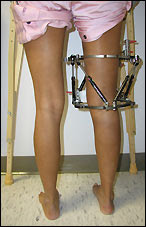
Causes
There are many causes of leg length discrepancy. Structural inequality is due to interference of normal bone growth of the lower extremity, which can occur from trauma or infection in a child. Functional inequality has many causes, including Poliomyelitis or other paralytic deformities can retard bone growth in children. Contracture of the Iliotibial band. Scoliosis or curvature of the spine. Fixed pelvic obliquity. Abduction or flexion contraction of the hip. Flexion contractures or other deformities of the knee. Foot deformities.
Symptoms
LLD do not have any pain or discomfort directly associated with the difference of one leg over the other leg. However, LLD will place stress on joints throughout the skeletal structure of the body and create discomfort as a byproduct of the LLD. Just as it is normal for your feet to vary slightly in size, a mild difference in leg length is normal, too. A more pronounced LLD, however, can create abnormalities when walking or running and adversely affect healthy balance and posture. Symptoms include a slight limp. Walking can even become stressful, requiring more effort and energy. Sometimes knee pain, hip pain and lower back pain develop. Foot mechanics are also affected causing a variety of complications in the foot, not the least, over pronating, metatarsalgia, bunions, hammer toes, instep pain, posterior tibial tendonitis, and many more.
Diagnosis
The only way to decipher between anatomical and functional leg length inequalities (you can have both) is by a physical measurement and series of biomechanical tests. It is actually a simple process and gets to the true cause of some runner?s chronic foot, knee, hip and back pain. After the muscles are tested and the legs are measured it may be necessary to get a special X-ray that measures both of your thighs (Femurs) and legs (Tibias). The X-ray is read by a medical radiologist who provides a report of the actual difference down to the micrometer leaving zero room for error. Once the difference in leg length is known, the solution becomes clear.
Non Surgical Treatment
Treatment for an LLD depends on the amount of difference and the cause, if known. The doctor will discuss treatment options carefully with you and your child before any decisions are made. It is important to note that treatment is planned with the child?s final height and leg lengths in mind, not the current leg lengths. Treatment is generally not needed if the child?s final LLD is predicted to be 2 centimeters or less at full height. However, the child should return to an orthopaedic doctor by age 10 for re-evaluation. Treatment is often recommended for LLDs predicted to be more than 2 centimeters at full height. If treatment is done, it usually doesn?t begin until the child starts walking. Possible treatment options include, A ?lift? in one shoe to level the child?s hips. This is often the only treatment needed for small discrepancies.

shoe lifts for men's shoes
Surgical Treatment
Large leg length inequalities can be treated by staged lengthenings or by simultaneous ipsilateral femoral and tibial lengthenings. Additionally, lengthenings can be combined with appropriately timed epiphysiodesis in an effort to produce leg length equality. Staged lengthenings are often used for congenital deficiencies such as fibular hemimelia, in which 15 cm or more may be needed to produce leg length equality. We typically plan for the final lengthening to be completed by age 13 or 14 years, and allow at least 3 years between lengthenings. Lengthening of both the tibia and femur simultaneously requires aggressive therapy and treatment of soft tissue contractures. Curran et al[57] reported the need for surgical release of soft tissue contractures in 3 of 8 patients treated with simultaneous ipsilateral femoral and tibial lengthenings. Lengthening over an IM nail can be done in an effort to decrease the amount of time the fixator needs to be worn and to prevent angular malalignment. This technique requires that the patient be skeletally mature and it carries a higher risk of osteomyelitis (up to 15%). Additionally, if premature consolidation occurs, a repeat corticotomy is more difficult.
Have you ever been told or noticed that one of your legs is a bit longer than the other? Do you have incidences of lower back pain? These two things could be related. Most individuals have a small difference in their leg lengths. For some, the discrepancy is small and negligible and will not be a contributor to lower back pain. This is usually the case for people if their leg length is less than 5 millimeters. However, a difference of leg lengths greater than 5 millimeters (1/4 inch) can contribute to lower back pain. If you have a leg length difference of greater than 9 mm, then you have a 6X greater likelihood of having an episode of lower back pain.
Causes
There are many causes of leg length discrepancy. Structural inequality is due to interference of normal bone growth of the lower extremity, which can occur from trauma or infection in a child. Functional inequality has many causes, including Poliomyelitis or other paralytic deformities can retard bone growth in children. Contracture of the Iliotibial band. Scoliosis or curvature of the spine. Fixed pelvic obliquity. Abduction or flexion contraction of the hip. Flexion contractures or other deformities of the knee. Foot deformities.
Symptoms
LLD do not have any pain or discomfort directly associated with the difference of one leg over the other leg. However, LLD will place stress on joints throughout the skeletal structure of the body and create discomfort as a byproduct of the LLD. Just as it is normal for your feet to vary slightly in size, a mild difference in leg length is normal, too. A more pronounced LLD, however, can create abnormalities when walking or running and adversely affect healthy balance and posture. Symptoms include a slight limp. Walking can even become stressful, requiring more effort and energy. Sometimes knee pain, hip pain and lower back pain develop. Foot mechanics are also affected causing a variety of complications in the foot, not the least, over pronating, metatarsalgia, bunions, hammer toes, instep pain, posterior tibial tendonitis, and many more.
Diagnosis
The only way to decipher between anatomical and functional leg length inequalities (you can have both) is by a physical measurement and series of biomechanical tests. It is actually a simple process and gets to the true cause of some runner?s chronic foot, knee, hip and back pain. After the muscles are tested and the legs are measured it may be necessary to get a special X-ray that measures both of your thighs (Femurs) and legs (Tibias). The X-ray is read by a medical radiologist who provides a report of the actual difference down to the micrometer leaving zero room for error. Once the difference in leg length is known, the solution becomes clear.
Non Surgical Treatment
Treatment for an LLD depends on the amount of difference and the cause, if known. The doctor will discuss treatment options carefully with you and your child before any decisions are made. It is important to note that treatment is planned with the child?s final height and leg lengths in mind, not the current leg lengths. Treatment is generally not needed if the child?s final LLD is predicted to be 2 centimeters or less at full height. However, the child should return to an orthopaedic doctor by age 10 for re-evaluation. Treatment is often recommended for LLDs predicted to be more than 2 centimeters at full height. If treatment is done, it usually doesn?t begin until the child starts walking. Possible treatment options include, A ?lift? in one shoe to level the child?s hips. This is often the only treatment needed for small discrepancies.
shoe lifts for men's shoes
Surgical Treatment
Large leg length inequalities can be treated by staged lengthenings or by simultaneous ipsilateral femoral and tibial lengthenings. Additionally, lengthenings can be combined with appropriately timed epiphysiodesis in an effort to produce leg length equality. Staged lengthenings are often used for congenital deficiencies such as fibular hemimelia, in which 15 cm or more may be needed to produce leg length equality. We typically plan for the final lengthening to be completed by age 13 or 14 years, and allow at least 3 years between lengthenings. Lengthening of both the tibia and femur simultaneously requires aggressive therapy and treatment of soft tissue contractures. Curran et al[57] reported the need for surgical release of soft tissue contractures in 3 of 8 patients treated with simultaneous ipsilateral femoral and tibial lengthenings. Lengthening over an IM nail can be done in an effort to decrease the amount of time the fixator needs to be worn and to prevent angular malalignment. This technique requires that the patient be skeletally mature and it carries a higher risk of osteomyelitis (up to 15%). Additionally, if premature consolidation occurs, a repeat corticotomy is more difficult.
Bunions Causes Symptoms And Treatments
Overview
Bunions are bony protrusions located at the base of the big toe that develop when the toe is slanted inward or overlaps the next toe. They can be very painful. Bunions form when the movement of the big toe influences the angle of the bones in the foot. The changes gradually develop into the characteristic bump, which over time becomes more and more noticeable.
Causes
The exact cause of bunions is unknown, but they tend to run in families. Wearing badly fitting shoes is thought to make bunions worse. It's also thought that bunions are more likely to occur in people with unusually flexible joints, which is why bunions sometimes occur in children. In some cases, certain health conditions, such as rheumatoid arthritis and gout, may also be responsible.
SymptomsThe most obvious symptoms of a bunion are. Pain in the area of the MTP joint, the joint where your big toe connects to your foot. Bending of the big toe in towards the other toes. An enlarged bump of bone or tissue at the MTP joint. Each symptom can range in degree from small to severe. Sometimes the pain can be sufficient to make it difficult to walk in normal shoes. Other symptoms may include. Swelling and inflammation of the skin around the MTP joint. Thickening of the skin in the area of the joint. Restricted motion in your big toe. Pressure from the inward bending of your big toe can affect your other toes, leading to corns on your smaller toes. Ingrown toenails on the smaller toes. Development of hammertoes in the other toes. Calluses on the bottom of your foot. If you have any of these symptoms, especially pain, displacement of your big toe or development of a bulge, you should consider consulting your physician. Even if you're not significantly bothered by some of these symptoms, bunions tend to continue getting bigger and more serious over time and should be taken care of before they do so.
Diagnosis
Your doctor will ask questions about your past health and carefully examine your toe and joint. Some of the questions might be: When did the bunions start? What activities or shoes make your bunions worse? Do any other joints hurt? The doctor will examine your toe and joint and check their range of motion. This is done while you are sitting and while you are standing so that the doctor can see the toe and joint at rest and while bearing weight. X-rays are often used to check for bone problems or to rule out other causes of pain and swelling. Other tests, such as blood tests or arthrocentesis (removal of fluid from a joint for testing), are sometimes done to check for other problems that can cause joint pain and swelling. These problems might include gout , rheumatoid arthritis , or joint infection.
Non Surgical Treatment
Bunions can be treated conservatively (without surgery) using simple measures such as well-fitting shoes, orthoses simple painkillers and padding. Physiotherapy can help improve associated muscle imbalances. Such measures will not correct or even stop the deformity but they can help with symptoms. When non-surgical treatments prove insufficient, surgery can relieve your pain, correct any related foot deformity and help you resume your normal activities.

Surgical Treatment
In 2010, the National Institute for Health and Care Excellence (NICE) published guidance about a minimally invasive surgical procedure to treat bunions. The aim of the procedure is to repair the tilting of the big toe. The technique can be carried out under a local anaesthetic or a general anaesthetic, using X-rays or an endoscope for guidance. The type of endoscope used will be a long, thin, rigid tube with a light source and video camera at one end. One or more incisions will be made near the big toe so that bone-cutting instruments can be inserted. These will be used to remove the bunion and to divide one or more bones located at the front of the foot. Wires, screws or plates will be used to keep the divided bones in place. After the procedure, you may need to wear a plaster cast or dressing to keep your foot in the correct position until the bones have healed. You may be given a special surgical shoe that enables you to walk on your heel. As the procedure is relatively new, there's little in the way of reliable evidence regarding its safety or effectiveness.
Prevention
Bunions often become painful if they are allowed to progress. But not all bunions progress. Many bunion problems can be managed without surgery. In general, bunions that are not painful do not need surgical correction. For this reason, orthopaedic surgeons do not recommend ?preventive? surgery for bunions that do not hurt, with proper preventive care, they may never become a problem.
Bunions are bony protrusions located at the base of the big toe that develop when the toe is slanted inward or overlaps the next toe. They can be very painful. Bunions form when the movement of the big toe influences the angle of the bones in the foot. The changes gradually develop into the characteristic bump, which over time becomes more and more noticeable.
Causes
The exact cause of bunions is unknown, but they tend to run in families. Wearing badly fitting shoes is thought to make bunions worse. It's also thought that bunions are more likely to occur in people with unusually flexible joints, which is why bunions sometimes occur in children. In some cases, certain health conditions, such as rheumatoid arthritis and gout, may also be responsible.
SymptomsThe most obvious symptoms of a bunion are. Pain in the area of the MTP joint, the joint where your big toe connects to your foot. Bending of the big toe in towards the other toes. An enlarged bump of bone or tissue at the MTP joint. Each symptom can range in degree from small to severe. Sometimes the pain can be sufficient to make it difficult to walk in normal shoes. Other symptoms may include. Swelling and inflammation of the skin around the MTP joint. Thickening of the skin in the area of the joint. Restricted motion in your big toe. Pressure from the inward bending of your big toe can affect your other toes, leading to corns on your smaller toes. Ingrown toenails on the smaller toes. Development of hammertoes in the other toes. Calluses on the bottom of your foot. If you have any of these symptoms, especially pain, displacement of your big toe or development of a bulge, you should consider consulting your physician. Even if you're not significantly bothered by some of these symptoms, bunions tend to continue getting bigger and more serious over time and should be taken care of before they do so.
Diagnosis
Your doctor will ask questions about your past health and carefully examine your toe and joint. Some of the questions might be: When did the bunions start? What activities or shoes make your bunions worse? Do any other joints hurt? The doctor will examine your toe and joint and check their range of motion. This is done while you are sitting and while you are standing so that the doctor can see the toe and joint at rest and while bearing weight. X-rays are often used to check for bone problems or to rule out other causes of pain and swelling. Other tests, such as blood tests or arthrocentesis (removal of fluid from a joint for testing), are sometimes done to check for other problems that can cause joint pain and swelling. These problems might include gout , rheumatoid arthritis , or joint infection.
Non Surgical Treatment
Bunions can be treated conservatively (without surgery) using simple measures such as well-fitting shoes, orthoses simple painkillers and padding. Physiotherapy can help improve associated muscle imbalances. Such measures will not correct or even stop the deformity but they can help with symptoms. When non-surgical treatments prove insufficient, surgery can relieve your pain, correct any related foot deformity and help you resume your normal activities.
Surgical Treatment
In 2010, the National Institute for Health and Care Excellence (NICE) published guidance about a minimally invasive surgical procedure to treat bunions. The aim of the procedure is to repair the tilting of the big toe. The technique can be carried out under a local anaesthetic or a general anaesthetic, using X-rays or an endoscope for guidance. The type of endoscope used will be a long, thin, rigid tube with a light source and video camera at one end. One or more incisions will be made near the big toe so that bone-cutting instruments can be inserted. These will be used to remove the bunion and to divide one or more bones located at the front of the foot. Wires, screws or plates will be used to keep the divided bones in place. After the procedure, you may need to wear a plaster cast or dressing to keep your foot in the correct position until the bones have healed. You may be given a special surgical shoe that enables you to walk on your heel. As the procedure is relatively new, there's little in the way of reliable evidence regarding its safety or effectiveness.
Prevention
Bunions often become painful if they are allowed to progress. But not all bunions progress. Many bunion problems can be managed without surgery. In general, bunions that are not painful do not need surgical correction. For this reason, orthopaedic surgeons do not recommend ?preventive? surgery for bunions that do not hurt, with proper preventive care, they may never become a problem.
Hallux Valgus Deformity
Overview
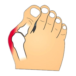
A bunion is an enlargement of ?the metatarsophalangeal (MTP) joint?, this is the big toe?s base joint that forms when the bone or tissue moves out of place. This forces the toe to bend toward the others, causing an often painful lump of bone on the foot. The MTP joint, which carries lots of weight, is stiff and sore, making even the wearing of shoes difficult or impossible. It also causes swelling of the feet to occur. Hammer toes are also associated with the formation of bunions.
Causes
Contributing factors may include excessive foot pronation, wearing tight and pointed-toe shoes, and occasionally trauma. Joint misalignment causes osteoarthritis with cartilage erosion and exostosis formation, resulting in joint motion being limited (hallux limitus) or eliminated (hallux rigidus). In late stages, synovitis occurs, causing joint swelling. In reaction to pressure from tight shoes, an adventitious bursa can develop medial to the joint prominence, which can become painful, swollen, and inflamed.
SymptomsIf you have a bunion, you may have pain or stiffness of your big toe joint, swelling of your big toe joint, difficulty walking, difficulty finding shoes that fit. These symptoms may be caused by conditions other than bunions, but if you have any of these symptoms, see your doctor.
Diagnosis
Before examining your foot, the doctor will ask you about the types of shoes you wear and how often you wear them. He or she also will ask if anyone else in your family has had bunions or if you have had any previous injury to the foot. In most cases, your doctor can diagnose a bunion just by examining your foot. During this exam, you will be asked to move your big toe up and down to see if you can move it as much as you should be able to. The doctor also will look for signs of redness and swelling and ask if the area is painful. Your doctor may want to order X-rays of the foot to check for other causes of pain, to determine whether there is significant arthritis and to see if the bones are aligned properly.
Non Surgical Treatment
The treatment of a bunion depends entirely on how uncomfortable it is. Realistically, there are only two ways to treat a bunion: either change the size and shape of the shoe or change the size and shape of the foot. Once a bunion gets to be irritating or painful and shoe wear is uncomfortable, surgery may be recommended.

Surgical Treatment
Most bunions can be treated without surgery. But when nonsurgical treatments are not enough, surgery can relieve your pain, correct any related foot deformity, and help you resume your normal activities. An orthopaedic surgeon can help you decide if surgery is the best option for you. Whether you?ve just begun exploring treatment for bunions or have already decided with your orthopaedic surgeon to have surgery, this booklet will help you understand more about this valuable procedure.
A bunion is an enlargement of ?the metatarsophalangeal (MTP) joint?, this is the big toe?s base joint that forms when the bone or tissue moves out of place. This forces the toe to bend toward the others, causing an often painful lump of bone on the foot. The MTP joint, which carries lots of weight, is stiff and sore, making even the wearing of shoes difficult or impossible. It also causes swelling of the feet to occur. Hammer toes are also associated with the formation of bunions.
Causes
Contributing factors may include excessive foot pronation, wearing tight and pointed-toe shoes, and occasionally trauma. Joint misalignment causes osteoarthritis with cartilage erosion and exostosis formation, resulting in joint motion being limited (hallux limitus) or eliminated (hallux rigidus). In late stages, synovitis occurs, causing joint swelling. In reaction to pressure from tight shoes, an adventitious bursa can develop medial to the joint prominence, which can become painful, swollen, and inflamed.
SymptomsIf you have a bunion, you may have pain or stiffness of your big toe joint, swelling of your big toe joint, difficulty walking, difficulty finding shoes that fit. These symptoms may be caused by conditions other than bunions, but if you have any of these symptoms, see your doctor.
Diagnosis
Before examining your foot, the doctor will ask you about the types of shoes you wear and how often you wear them. He or she also will ask if anyone else in your family has had bunions or if you have had any previous injury to the foot. In most cases, your doctor can diagnose a bunion just by examining your foot. During this exam, you will be asked to move your big toe up and down to see if you can move it as much as you should be able to. The doctor also will look for signs of redness and swelling and ask if the area is painful. Your doctor may want to order X-rays of the foot to check for other causes of pain, to determine whether there is significant arthritis and to see if the bones are aligned properly.
Non Surgical Treatment
The treatment of a bunion depends entirely on how uncomfortable it is. Realistically, there are only two ways to treat a bunion: either change the size and shape of the shoe or change the size and shape of the foot. Once a bunion gets to be irritating or painful and shoe wear is uncomfortable, surgery may be recommended.
Surgical Treatment
Most bunions can be treated without surgery. But when nonsurgical treatments are not enough, surgery can relieve your pain, correct any related foot deformity, and help you resume your normal activities. An orthopaedic surgeon can help you decide if surgery is the best option for you. Whether you?ve just begun exploring treatment for bunions or have already decided with your orthopaedic surgeon to have surgery, this booklet will help you understand more about this valuable procedure.
Hallux Abducto Valgus Correction Exercises
Overview

A bunion is an enlargement at the base of the big toe caused by a misalignment of the joint. Hallux valgus or hallux abducto valgus (HAV) is the name used for the deviated position. of the big toe and a bunion refers to the enlargement of the joint, most of the time the two go together and can just be referred to as ?bunions?. Bunions are really only a symptom of faulty foot mechanics and are usually caused by the foot we inherit and inappropriate footwear use. As the big toe bends towards the others this lump becomes larger and the bunion can become painful - arthritis and stiffness can eventually develop.
Causes
The commonest cause of bunions is prolonged wearing of poorly designed shoes such as the narrow high heels that women wear. This is one of the reasons why bunions are much more common in women than in men. There is also a hereditary component to bunions in that many times we will see a grandmother, mother and daughter all with various stages of bunions. 38% of women in the United States wear shoes that are too small and 55% of women have some degree of bunion formation. Bunions are 9 times more common in women than they are in men.
SymptomsRedness, swelling, or pain along the inside margin of the foot just behind the great toe. Moderate to severe discomfort at the bunion when wearing shoes, particularly if tight fitting. A painful callus may develop over the bunion. Sometimes a painful corn on the adjacent sides of the first and second toes. Irritation if there is overlapping of the first and second toes. Arthritis may cause stiffness and discomfort in the joint between the great toe and the first metatarsal. There may be a fluid filled cyst or bursa between the skin and the "bunion bone". Skin over the bunion may break down causing an ulceration, which can become infected.
Diagnosis
Your family doctor or chiropodist /podiatrist can identify a bunion simply by examining your foot. During the exam, your big toe will be moved up and down to determine if your range of motion is limited. You will be examined for signs of redness or swelling and be questioned about your history of pain. A foot x-ray can show an abnormal angle between the big toe and the foot. In some cases, arthritis may also be seen. A X-ray of your foot may help identify the cause of the bunion and rate its severity.
Non Surgical Treatment
Initial treatment of bunions may include wearing comfortable, well-fitting footwear (particularly shoes that conform to the shape of the foot and do not cause pressure areas) or the use of splints and orthotics (special shoe inserts shaped to your feet) to reposition the big toe. For bunions caused by arthritis, medications may help reduce pain and swelling. If nonsurgical treatment fails, your doctor may suggest surgery, which resolves the problem in nearly all persons. The goal of surgery is to relieve pain and correct as much deformity as possible. The surgery is not cosmetic and is not meant to improve the appearance of the foot. Other related procedures that may be used to help diagnose foot disorders include X-rays of the bone and foot.

Surgical Treatment
Bunions can cause pain and difficulty wearing certain shoes. When simple treatments don't relieve your symptoms, surgery may be considered for treatment of the bunion. What are the signs that surgery may be the right treatment for your bunion? In general, surgery is recommended only when pain from the bunion prevents a patient from wearing normal shoes. There is a common misconception that surgical treatments for a bunion are better and quicker than non-surgical treatments. Unfortunately, patients who rush into surgery may have unrealistic expectations, and may be unsatisfied with surgery.
Prevention
Proper footwear may prevent bunions. Wear roomy shoes that have wide and deep toe boxes (the area that surrounds the toes), low or flat heels, and good arch supports. Avoid tight, narrow, or high-heeled shoes that put pressure on the big toe joint. Medicine will not prevent or cure bunions.
A bunion is an enlargement at the base of the big toe caused by a misalignment of the joint. Hallux valgus or hallux abducto valgus (HAV) is the name used for the deviated position. of the big toe and a bunion refers to the enlargement of the joint, most of the time the two go together and can just be referred to as ?bunions?. Bunions are really only a symptom of faulty foot mechanics and are usually caused by the foot we inherit and inappropriate footwear use. As the big toe bends towards the others this lump becomes larger and the bunion can become painful - arthritis and stiffness can eventually develop.
Causes
The commonest cause of bunions is prolonged wearing of poorly designed shoes such as the narrow high heels that women wear. This is one of the reasons why bunions are much more common in women than in men. There is also a hereditary component to bunions in that many times we will see a grandmother, mother and daughter all with various stages of bunions. 38% of women in the United States wear shoes that are too small and 55% of women have some degree of bunion formation. Bunions are 9 times more common in women than they are in men.
SymptomsRedness, swelling, or pain along the inside margin of the foot just behind the great toe. Moderate to severe discomfort at the bunion when wearing shoes, particularly if tight fitting. A painful callus may develop over the bunion. Sometimes a painful corn on the adjacent sides of the first and second toes. Irritation if there is overlapping of the first and second toes. Arthritis may cause stiffness and discomfort in the joint between the great toe and the first metatarsal. There may be a fluid filled cyst or bursa between the skin and the "bunion bone". Skin over the bunion may break down causing an ulceration, which can become infected.
Diagnosis
Your family doctor or chiropodist /podiatrist can identify a bunion simply by examining your foot. During the exam, your big toe will be moved up and down to determine if your range of motion is limited. You will be examined for signs of redness or swelling and be questioned about your history of pain. A foot x-ray can show an abnormal angle between the big toe and the foot. In some cases, arthritis may also be seen. A X-ray of your foot may help identify the cause of the bunion and rate its severity.
Non Surgical Treatment
Initial treatment of bunions may include wearing comfortable, well-fitting footwear (particularly shoes that conform to the shape of the foot and do not cause pressure areas) or the use of splints and orthotics (special shoe inserts shaped to your feet) to reposition the big toe. For bunions caused by arthritis, medications may help reduce pain and swelling. If nonsurgical treatment fails, your doctor may suggest surgery, which resolves the problem in nearly all persons. The goal of surgery is to relieve pain and correct as much deformity as possible. The surgery is not cosmetic and is not meant to improve the appearance of the foot. Other related procedures that may be used to help diagnose foot disorders include X-rays of the bone and foot.
Surgical Treatment
Bunions can cause pain and difficulty wearing certain shoes. When simple treatments don't relieve your symptoms, surgery may be considered for treatment of the bunion. What are the signs that surgery may be the right treatment for your bunion? In general, surgery is recommended only when pain from the bunion prevents a patient from wearing normal shoes. There is a common misconception that surgical treatments for a bunion are better and quicker than non-surgical treatments. Unfortunately, patients who rush into surgery may have unrealistic expectations, and may be unsatisfied with surgery.
Prevention
Proper footwear may prevent bunions. Wear roomy shoes that have wide and deep toe boxes (the area that surrounds the toes), low or flat heels, and good arch supports. Avoid tight, narrow, or high-heeled shoes that put pressure on the big toe joint. Medicine will not prevent or cure bunions.
A Secrets And Techniques Measuring Feet
No matter how young you are, as soon as you have miscalculated a step or two then your foot pain walking problems will surely present themselves. There are also times when severe pain, especially at the ankle joint, is brought about by wearing impossibly high heels for a considerable period. The pain you are experiencing is not focused on one part of the foot since the foot has several ligaments, muscles, tendons and joints that once you have injured one element, the entire foot will feel pain.
The most common symptoms of foot Bunions would be swelling as well as redness on the toe joint area. There is also the presence of a bulge along the outside of the toe. The skin on the toe area is also present on the big toe. You also develop calluses and corns and these can happen when two of the toes overlap each other. The conditions also produces considerable amount of pain and discomfort. Lastly, the big toe becomes restricted in movements. The pain that the bunions cause can be so severe that waling on regular shoes is impossible and you have to immediately sit down and just wait for the pain to subside. If the pain has become persistent and the shoes you are wearing are not fitting you anymore, it is necessary to talk to your doctor about foot .

Once a bunion begins, the pain and frustration alone are enough to send you running into the office of the closest foot surgeon. The bulging of the Halux Valgus begins to change the shape of one of your feet, making it difficult to find comfortable shoes. Without a shoe on, the toes are crowded; when you try to put any type of shoe on, the crowding is worse and the pain is often unbearable. There are not a lot of place that a person can go without wearing shoes.
There are many kinds of foot problems that can be corrected by minor orthopedic footwear. Some of these problems include painful heels, tired feet, flat feet, bunions, calluses, corns, ingrown or thick toe nails, athlete's foot, dry foot, warts, hammer toes and diabetic foot problems.
You should always consult a doctor for treatment of gout. But some of the drugs used for gout - such as allopurinol, colchicine, indomethacin, and prednisone - can cause serious side effects.
The toes are often injured by ill-fitting shoes. Both the forefoot and the hindfoot absorb tons of force each and every day. They also contain dozens of bones on which bone spurs may form over time. A bone spur is an overgrowth of bone that can cause discomfort and pain. They are the most common cause of Foot Pain.
HEALTHY HIP MECHANICS - The inner thighs stabilize the leg as it swings forward and backwards for a healthy stride to walk and run. They also are critical for support and movement for lateral (side-to-side) movement. Sports like basketball, tennis, racquetball and horseback riding would be impossible without strong inner thighs.
Unfortunately, a lot of the things that go wrong with your feet either require surgery or it is so little that nothing can be done and it just has to heal on its own. Some things can be prevented, but other things just happen some times. You walk around every day all day and there is a lot of room for something to go wrong. If you play sports, it is even easier to get hurt. Being careful will prevent a lot of issues but if problems occur and sometimes they just do, going to the doctor would be the best bet even if you think it is just something small. It could be something more serious that needs attention.